


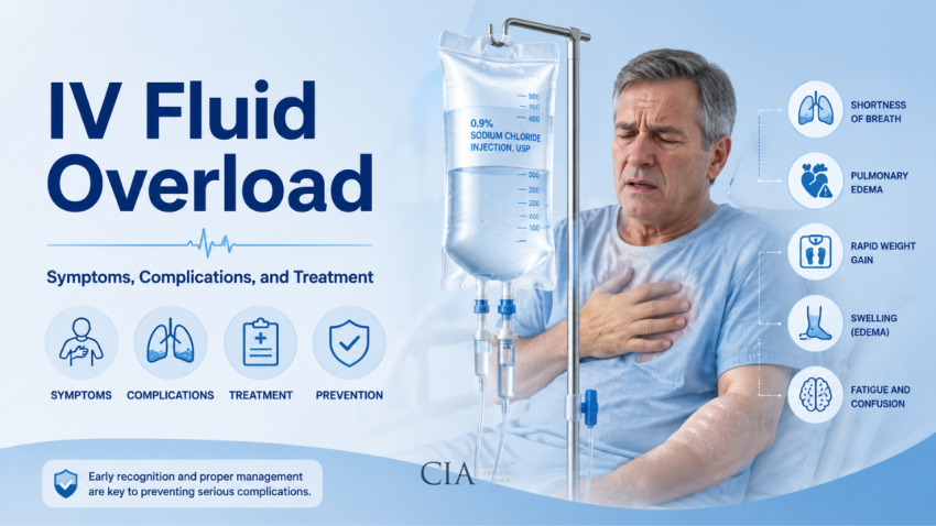
IV Fluid Overload: Symptoms, Complications, and Treatment
IV fluid overload, also called volume overload or iatrogenic hypervolemia, is a well-documented complication of intravenous fluid therapy, particularly in critical care settings. It is not rare.
Studies of mechanically ventilated ICU patients show that 50 to 70% develop a significantly positive fluid balance during their stay, and that cumulative positive balance is independently associated with worse outcomes: higher mortality, prolonged mechanical ventilation, and longer ICU and hospital stays.
Clinicians managing IV therapy need to recognize the early signs, understand which patients are most vulnerable, and know the evidence-based interventions for prevention and treatment.
What Is IV Fluid Overload?
IV fluid overload occurs when the volume of fluid administered exceeds the body’s capacity to excrete or redistribute it. The result is expansion of the extracellular fluid compartment, both intravascular and interstitial, leading to edema, impaired organ function, and potentially life-threatening complications.
Fluid overload is most commonly assessed through fluid balance calculations: total fluid input (IV fluids, enteral feeds, medications) minus total output (urine, drains, insensible losses). A cumulative positive balance greater than 10% of body weight is associated with significantly worse clinical outcomes in critically ill patients. Weight gain of more than 1 kg per day is a practical bedside indicator of fluid retention.
The pathophysiology is not simply a plumbing problem. Excess isotonic crystalloid (particularly 0.9% normal saline) can cause dilutional hyperchloremic metabolic acidosis, worsen endothelial glycocalyx degradation, impair cardiac function, and promote systemic inflammatory cascades that amplify organ dysfunction.
What Are the Symptoms of IV Fluid Overload?
Symptoms appear across multiple organ systems and can range from subtle early signs to acute respiratory failure. Recognition depends on systematic assessment.
Pulmonary Symptoms
Pulmonary manifestations are often the most clinically urgent. Excess fluid shifts into the interstitial and alveolar spaces of the lungs, increasing work of breathing and impairing gas exchange.
- Dyspnea and increased work of breathing, often the earliest patient-reported symptom
- Tachypnea, respiratory rate above 20 breaths per minute
- Crackles (rales) on auscultation, bilateral basal crackles indicate interstitial or alveolar fluid
- Decreased oxygen saturation on pulse oximetry
- Frothy or pink-tinged sputum, indicates frank pulmonary edema, a late and dangerous sign
- Orthopnea, inability to lie flat without dyspnea, a hallmark of cardiogenic pulmonary congestion
Cardiovascular Symptoms
The cardiovascular system bears the increased preload directly. In patients with preserved cardiac function, the heart may compensate temporarily; in those with reduced ejection fraction or valvular disease, decompensation occurs rapidly.
- Elevated jugular venous pressure (JVP), distension visible at 45 degrees indicates right-sided congestion
- S3 gallop on cardiac auscultation, low-frequency third heart sound reflecting impaired ventricular filling
- Tachycardia, compensatory response to reduced stroke volume
- Hypertension, in early overload as cardiac output increases; may shift to hypotension with decompensation
- BNP (B-type natriuretic peptide) elevation, BNP greater than 100 pg/mL is associated with volume overload; levels above 400 pg/mL suggest significant cardiac stress
Systemic and Peripheral Symptoms
Fluid distribution beyond the intravascular space produces characteristic peripheral findings.
- Pitting edema, graded I to IV; begins at dependent areas (ankles, sacrum in supine patients) and ascends with severity
- Facial or periorbital edema, common in hypoalbuminemic patients and neonates
- Ascites, abdominal fluid accumulation, particularly in cirrhotic patients
- Weight gain, a 1 kg increase equates to approximately 1 liter of retained fluid; daily weights are a sensitive early monitoring tool
- Decreased urine output, prerenal physiology as the kidneys respond to perceived low effective circulating volume despite actual volume excess
- Dilutional hyponatremia, serum sodium below 135 mEq/L, most common with hypotonic fluid infusions or large-volume 5% dextrose
How Is IV Fluid Overload Diagnosed?
Diagnosis is clinical, supported by targeted investigations. No single test is definitive.
Physical examination findings, bilateral crackles, elevated JVP, peripheral edema, and rapid weight gain, provide the initial picture. Daily weights are the most sensitive bedside tool; weight trends are more informative than single measurements.
Laboratory markers: BNP or NT-proBNP for cardiac volume stress; serum sodium for dilutional changes; serum chloride and bicarbonate to detect hyperchloremic acidosis from excess normal saline; albumin level (hypoalbuminemia lowers oncotic pressure and worsens edema). Serum creatinine and urine output trends reflect renal impact.
Imaging: Chest X-ray showing pulmonary vascular congestion, Kerley B lines, or frank alveolar edema confirms pulmonary overload. Point-of-care lung ultrasound (B-lines or lung rockets) is increasingly used at the bedside and is more sensitive than CXR for early interstitial edema. Inferior vena cava (IVC) ultrasound assessing collapsibility can estimate right atrial pressure, though accuracy varies.
What Are the Complications of IV Fluid Overload?
The consequences extend well beyond the lungs. Cumulative fluid overload drives complications in nearly every organ system.
- Acute Respiratory Distress Syndrome (ARDS), fluid overload worsens oxygenation in existing ARDS and can precipitate it in susceptible patients
- Prolonged mechanical ventilation, the FACTT trial (NEJM, 2006) demonstrated that a conservative fluid strategy reduced ventilator-free days by 2.5 days in ARDS patients compared to a liberal strategy
- Acute kidney injury, abdominal compartment syndrome secondary to bowel edema increases intra-abdominal pressure, reducing renal perfusion pressure
- Abdominal compartment syndrome, intra-abdominal pressure above 20 mmHg with associated organ dysfunction; associated with massive fluid resuscitation
- Wound dehiscence and impaired healing, interstitial edema reduces tissue oxygen delivery and nutrient diffusion
- Ileus and bowel edema, impairs enteral nutrition initiation and recovery of GI function
- Electrolyte disturbances, hyperchloremia, hyponatremia, hypokalemia (with diuresis)
- Increased mortality, multiple observational studies and the FACTT trial data link sustained positive fluid balance to higher 60-day mortality in critically ill patients
How Is IV Fluid Overload Treated?
Treatment centers on removing excess fluid while maintaining adequate organ perfusion, a balance that requires careful clinical judgment.
Diuresis is the primary intervention for most patients with intact renal function. Furosemide (a loop diuretic) is first-line: typical starting doses are 20 to 40 mg IV, with titration based on urine output response. The target is a urine output of 0.5 to 1.0 mL/kg/hour during active de-resuscitation. Patients on chronic furosemide often require higher doses due to tolerance; torsemide or bumetanide may be used in diuretic resistance.
For patients with diuretic resistance or significant AKI, continuous renal replacement therapy (CRRT) or intermittent hemodialysis with ultrafiltration can achieve fluid removal at controlled rates. This is particularly relevant in anuric or oliguric patients.
Fluid restriction, reducing or stopping non-essential IV infusions, converting oral medications, and limiting flush volumes, is a simple but often overlooked step in early overload management.
Albumin infusion is sometimes used in hypoalbuminemic patients to improve oncotic pressure and mobilize interstitial fluid back into the intravascular space, though evidence for mortality benefit is mixed.
Oxygen supplementation and positioning, elevating the head of the bed, high-flow nasal cannula, or non-invasive positive pressure ventilation, address pulmonary symptoms while diuresis is initiated.
How Can IV Fluid Overload Be Prevented?
Prevention begins with intentional fluid prescribing. Each liter of IV fluid should have a specific indication, a defined rate and volume, and a reassessment interval, the same rigor applied to any drug order.
Goal-directed fluid therapy (GDFT) protocols use dynamic hemodynamic parameters, stroke volume variation (SVV), pulse pressure variation (PPV), or response to fluid bolus challenges, to guide fluid administration. This approach is well-validated in the perioperative setting and has demonstrated reductions in complications compared to static protocol-based infusion.
The SMART trial (NEJM, 2018) compared balanced crystalloids (lactated Ringer’s or Plasma-Lyte) to 0.9% normal saline in ICU patients and found a small but significant reduction in the composite of death, new renal replacement therapy, or persistent renal dysfunction in the balanced crystalloid group. Fluid choice affects outcomes; saline-induced hyperchloremic acidosis impairs renal perfusion and worsens fluid balance.
Daily reassessment of each IV line and infusion should be standard practice. Fluids that no longer have a clear indication should be discontinued.
Which Patients Are at Highest Risk for IV Fluid Overload?
The following patient populations are at substantially higher risk and require more conservative fluid strategies and earlier monitoring:
- Congestive heart failure (CHF), reduced ejection fraction limits the heart’s ability to handle even modest increases in preload; even 500 mL boluses can precipitate acute decompensation
- Chronic kidney disease (CKD) and end-stage renal disease, impaired ability to excrete fluid loads; accelerated shift to overload
- Cirrhosis and hepatic failure, hypoalbuminemia and portal hypertension make these patients exceptionally prone to ascites and peripheral edema with fluid loading
- Elderly patients, reduced cardiac reserve, decreased renal function, and lower serum albumin all increase vulnerability
- Post-operative patients, particularly after major abdominal or cardiac surgery, where third-spacing and capillary leak are significant
- Sepsis patients in the fluid de-escalation phase, initial resuscitation may require large volumes, but failure to de-escalate once hemodynamics are restored is a common driver of ICU fluid overload
- ARDS patients, the FACTT data is unambiguous: conservative fluid management improves clinical outcomes compared to liberal fluid strategies in established ARDS
References
- https://pubmed.ncbi.nlm.nih.gov/19436332/
- https://pubmed.ncbi.nlm.nih.gov/33009098/
- https://pubmed.ncbi.nlm.nih.gov/10319771/
- https://pubmed.ncbi.nlm.nih.gov/29485925/
- https://pubmed.ncbi.nlm.nih.gov/35504747/
- https://pubmed.ncbi.nlm.nih.gov/27738801/
- https://pmc.ncbi.nlm.nih.gov/articles/PMC6821102/
- https://pmc.ncbi.nlm.nih.gov/articles/PMC1919391/
- https://www.nejm.org/doi/full/10.1056/NEJMoa062200
- https://www.nejm.org/doi/full/10.1056/NEJMoa1711584
- https://link.springer.com/article/10.1007/s00540-016-2261-7

